Getting to the root of soil parasite infections in wheat
School of Agriculture, Food and Wine researchers have uncovered how a common parasite affects wheat crops by applying new technology to address an old problem.
Using high-resolution 3D imaging, PhD student Kara Levin discovered how cereal cyst nematodes interfere with plant root development to hijack water and nutrient flow.
Cyst nematodes are soil-borne pests that infect plant roots. In southern Australia, cereal cyst nematodes (CCN) once caused devastating yield losses in wheat and barley crops. Over many years, researchers at the University of Adelaide and SARDI guided efforts to bring these parasites under control. Despite this, it remained uncertain exactly how these nematodes interfere with root development and crop yield.
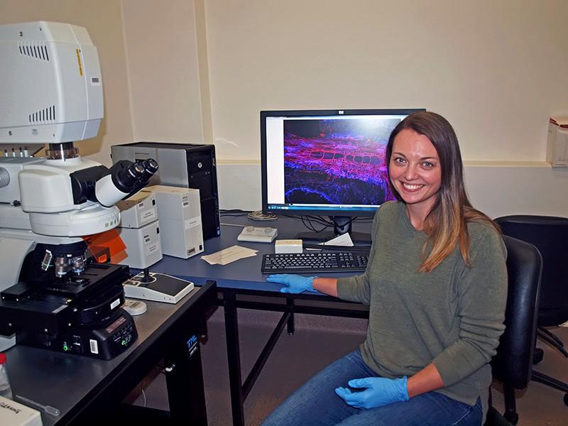
Kara Levin used confocal microscopy to generate 3D images of cereal cyst nematode infection in wheat roots.
New research, published in the journal Scientific Reports, used cutting edge imaging technology to take a close look within CCN-infected wheat roots.
“By combining new techniques from plant biology, microscopy and computer modelling, we were able to look deep within CCN-infected wheat roots to generate 3D models that show nematodes and their effects on plant tissues.” said lead author Kara Levin from the School of Agriculture, Food and Wine, and the Waite Research Institute.This revealed that CCN infection dramatically affects the development of xylem vessels that transport water and mineral nutrients from the root to the rest of the plant.
Above: Rotating 3-D model of a nematode infection site within a wheat root. The nematode (shown in red) and its feeding site (shown in blue) are positioned next to the water transport system of the roots (shown in green and yellow, green being parts of the root invaded by the feeding site)
“Although microscopy has been used to investigate this system for decades, this study is the first to show that xylem cells in infected roots become short and fat ‘bubbles’ rather than long and narrow ‘tubes’. This is likely to impede water transport within the roots.” Kara Levin - School of Agriculture, Food and Wine
“Although microscopy has been used to investigate this system for decades, this study is the first to show that xylem cells in infected roots become short and fat ‘bubbles’ rather than long and narrow ‘tubes’. This is likely to impede water transport within the roots.”
“We also saw clear evidence of nematode feeding sites having ‘invaded’ xylem vessels.”
Plant breeding and genetics expert Professor Diane Mather said that this new knowledge is now being applied to investigate how some varieties are able to resist this parasite.
“This research means we now have a clearer picture of how nematode infections affect root development, which is important in determining resistance mechanisms in wheat that could be exploited in breeding programs.”
“The innovative use of imaging techniques can also be applied to other plant species and other research questions.”